Showing posts with label CVS. Show all posts

Monday, May 11, 2009

thrills

0 comments
Mr. Jo, 45 years old man with previous history of right coronary artery infarction presented to HUSM. He has progressive effort intolerance and abdominal discomfort. On abdominal examination, there are positive fluid thrills and shifting dullness.
  • What is the sign of the abdominal findings in this patient and state the possible cause. (1m)
  • What are the 4 expected clinical signs that might be obtained in this patient? (2m)
  • Outline the pathophysiology of the abdominal signs. (2m)


Wassalam.

Friday, May 8, 2009

drug user and murmur... are they related?

0 comments

18 years old man who is an intravenous drug user came to HUSM complaining of having fever and dark colored urine. Physical examination revealed that he has low grade fever, hematuria, clubbing, and subconjuctival hemorrhage. Murmur was also auscultated on tricuspid area.
  • What is the provisional diagnosis for this patient? (1m)
  • Outline the pathophysiology of hematuria in this patient. (4m)
  • What is the reason for the presence of murmur in this patient? (2m)
  • State 2 possible complications that might occur in this patient. (2m)


Wassalam.

Wednesday, March 25, 2009

ACS

0 comments
Ischemic Heart Disease can be divided into 2 categories:
1) Acute IHD / ACS - Unstable angina, NSTEMI, STEMI
2) Chronic IHD - Stable angina, Printzmetal angina


ACUTE IHD

1) Unstable Angina - No ST elevation

Pain

- more persistent and severe course
- characterized by at least 1 of these:
a) at rest / minimal exertion, lasts more than 20 minutes (if not interrupted by GTN)
b) described as frank pain & new onset (within 1 month)
c) more severe, prolong or frequent than the previous episode

2) NSTEMI (Subendocardial infarction) -No ST elevation
3) STEMI (Transmural infarction) - Have ST elevation /Q wave

How the chest pain is dexcribed for 2 & 3? - Use LORDSANFARO [but info here might not be fulfilling all these]

L- Substernal
O- Abrupt onset
R- To left arm, neck, jaw, or other chest areas.
D- More prolong (more than 20-30 minutes)
S- ?
A- ?
N- Burning in nature [might be confused with GERD]
F- ?
A- ?
R- Not relieved by rest and GTN.
O- ?

** ACS - is used to describe a spectrum of IHD that includes Unstable Angina, NSTEMI, STEMI.


So, how to diagnose?
1) Do ECG - Any ST elevation? - If yes: STEMI, If no: Unstable angina and NSTEMI


How to differentiate Unstable angine & NSTEMI?
1) Do cardiac enzymes? If -ve: Unstable Angina

** LDH: "Last Diagnostic Hint" - which means the last enzyme.


Refer to: Pathophysio LWW [most] & internet.

that's all till now.


sharing is caring 12 :)


Wassalam.

Monday, March 23, 2009

identification data - shortness of breath,cough,frothy sputum

1 comments
Soalan 3
a 63 year old lady presented with shortness of breath and cough with frothy sputum.

On examination she has displaced heart beat and bibasal crepitations. She was diagnosed as a hypertensive since 15 years ago but defaulted treatment.

You are shown her chest radiograph

sate 2 abnormalities seen in the radiograph

Loss of costophrenic angle
Increase cardio-thoracic ratio (cardiomegaly)
Increase pulmonary vascular markings


State the most likely diagnosis

Pulmonary oedema

Briefly explain how hypertension lead to this patient’s condition

Hypertension → increase systemic pressure → heart need to pump harder to overcome the resistance → left heart failure → congested flow → extravasation of pulmonary vessels → pulmonary oedema

State 2 complication to diagnosis above

End organ failure (sbb heart cannot pump out blood, tak cukup blood supply keorgan2, organ failure)
Cardiogenic shock

-----------------------------------------------------------------------------------------
p/s: gmbar xray ni bukan 100% sama tp lebih kurang sama dgn id trial hr tu.
maaf sbb lambat post

jvp

0 comments
Questions

1. What does the Jugular Venous Pressure (JVP) tell us?
2. What is the position of the patient when JVP is done?
3. Give 3 causes for the elevated JVP.
4. Label a, c, x, v and y.
5. What condition might cause abnormal in a, x, and y?


Answers

1. JVP tells us about the right atrial and right ventricular function.

2. Patient should lies in supine position propped up 45 degrees.

3. 3 causes for the elevated JVP (choose 3):
a) Right ventricular failure
b) Tricuspid stenosis or regurgitation
c) Pericardial effusion or constrictive pericarditis
d) SVC obstruction
e) Fluid overload
f) Hyperdynamic circulation

4 & 5:


sharing is caring 10 :)

Wassalam.

Identification Data-Chest pain

2 comments
A 40 year old, male, teacher came to the casualty department complaining of chest pain, sudden onset radiating to left shoulder and jaw. He also had a history of being unable to lie flat and shortness of breath after walking for a few steps.


a. State one (1) abnormality of the ST segment shown in the ECG recording
St elevation- lead III, aVF, V3, V4, V5
ST depression- aVL

b. State one (1) other abnormality of the ECG recording shown
Deep Q wave at lead III

c. State the most likely diagnosis
Acute Myocardial Infarction

d. List two (2) other relevant investigations you would do and state your reason

Plasma biochemical marker - to detect any rise in the plasma concentration of enzyme and protein that are normally concentrated within cardiac cells. (in MI- rise in CK-MB, Troponin T and I)

Echocardiography- to check cardiac function and detecting important complication (e.g mural thrombus, cardiac rupture, VSD, mitral regurgitation, pericardial effusion)

~living well or living hell? Fighting for future!~

Thursday, March 19, 2009

SILHOUETTE? :)

0 comments

CARDIAC SILHOUETTE

  1. Right Atrium
  2. Right Ventricle
  3. Apex of Left Ventricle
  4. Superior Vena Cava
  5. Inferior Vena Cava
  6. Tricuspid Valve
  7. Pulmonary Valve
  8. Pulmonary Trunk
  9. Right Pulmonary Artery
  10. Left Pulmonary Artery

p/s: i don't think we've got to know all these in great details for now. but after all, SHARING IS CARING :)

sharing is caring 4 :)

Wassalam.

Sunday, March 15, 2009

ecg class

0 comments
by adam

9 steps for ecg interpretation

  1. cardiac axis
  2. heart rate
  3. rhythm
  4. P wave
  5. PR interval
  6. QRS complex
  7. ST segment
  8. T wave
  9. other wave-U wave can be normal

short notes

p/s;all ecg notes were prepared by adam,thanks=p

short notes on ECG

0 comments
right ventricular hypertrophy
  1. tall R wave in V1
  2. T inversion V1-V3/V4
  3. deep S in V6
  4. right axis
  5. RBBB

left ventricular hypertrophy

1) R wave in V6 greater than 25mm
2) R wave in V6 plus S in V1 greater than 35mm
3) Inverted T in V1 ,VL, V5-V6

left atrial hypertrophy

1) Bifid P wave

right atrial hypertrophy

1) Peaked P wave

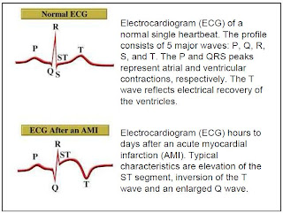
myocardial infarction

1) Normal ECG
2) Raised ST segment
3) Appearance of Q wave
4) Normalisation of ST segment
5) Invertion of T wave

*Site of infarction

Þ Anterior infarction – changes in V2 – V5
Þ Inferior infarction – changes in III and aVF
Þ Lateral infarction – changes in I, aVL, V6
Þ True posterior infarction – dominant R in V1

ecg-4

0 comments

st elevation in V1-V5 indicate ant MI

ST segment

  • normal-isoelectric
  • Elevation(like picture above)-Myocardial infarction/pericarditis
  • depression-myocardial ischemia

in pericarditis,all leads show ST elevation,but in MI,only respective leads correspond to that area shows ST elevation.

T wave

  • tall-hyperkalaemia
  • inverted-hypokalaemia

ecg-3

0 comments

rhythm

normal aka sinus rhythm

  • P wave precedes QRS wave
  • therefore,num of P wave equals with QRS wave
  • PR intervals-normal

sinus rhythm

sinus tachycardia

  • rate:fast(300/3=100 beats per minute)
  • the rest normal

    sinus tachycardia

P wave

  • wide and bifid P wave-in left atrial hypertrophy(might be due to mitral stenosis)
  • peak/tall P wave-in right atrial hypertrophy(might be due to tricuspid valve stenosis)
QRS wave
normal
i.its duration is no greater than 0.12 sec
ii.In a right ventricular lead (V1) the S wave is greater than the R wave
iii.In left ventricular leads (V5 or V6) the height of R wave is less than 25mm
iv.Left ventricular leads may show Q waves due to septal depolarization, but these are less than 1mm across and less than 2mm deep
v.R in V6 plus S in V1 less than 35mm
right ventricular hypertrophy
-tall R wave in V1 and deep S-wave in V6
left ventricular hypertrophy
-deep S wave in V1 and tall R wave in V6


ecg-2

0 comments
cardiac axis

The average direction of spread of the depolariation wave through the ventricle as seen from the frontal plane

determine by the lead I,II,II->look at the QRS wave

right axis deviation
I-negative
II,III-positive

left axis deviation
I-positive
II,III-negative


u can also determine the cardiac axis by looking at the lead I and aVF.if patient is having left axis deviation,then check lead II.

ecg-1

0 comments

recall the normal ECG
  • P wave-depolarization of atrium (time-<0.12s,amplitude-0.5-2.5mm)
  • QRS wave-depolarization of ventricle(time-<0.12s,amplitude-1.2-20mm)
  • T wave-repolarization of ventricle(time-0.1-0.25s,amplitude<5mm)
  • PR interval-time from depolarization of atrium to the ventricle(0.12-0.2s)
  • QT interval-ventricular activity(depolarization n repolarization)
  • ST segment-time between end of ventricular depolarization and beginning of repolarization(time<0.2s)

normal heart rate:60-100 beats per minute
count from ECG;from R-R
  • 1500/num of small box between R-R
  • 300/num of large box between R-R

each small box-0.04 seconds,each large box-0.2 seconds.therefore,in 60 seconds,there are 1500 small boxes,and 300 large boxes.

from the ecg,between R-R,there are 5 large boxes.therefore,the heart rate is 300/5=60 beats per minute

if patient is having irregular rhythm,count the num of R-R interval in 30 large boxes,then times 10,the result is the heart rate.

p/s:part seterusnya akan dipostkan nanti,nak cari gmbr ecg dulu.khatam basic ye=p

-adam al anas-

Friday, March 13, 2009

modified jones criteria [only for major ones]

0 comments
Major criteria would be JONES itself

J- Joints (Migratiry polyarthritis)
O- O [imagine heart-shaped O] (Carditis)
N- Nodules (Subcutaneous Nodules)
E- Erythema Marginatum
S- Sydenham's criteria

or

CASES

C- Carditis
A- Arthritis
S- Sydenham's chorea
E- Erythema marginatum
S- Subcutaneous nodules

Choose which ever you prefer!!! (^_^)

Thursday, March 12, 2009

Duke's Criteria

0 comments
BE FEVEER
(*BE - Bacterial Endocarditis)

Major:
B = blood culture +ve >2 times 12 hr part
E = Endocardial involvement from Echo

Minor:
F = Fever
E = Echo findings (not fulfilling a major)
V = Vascular findings [Janeway lesion]
E = Evidences from microbiologic
E = Evidences from immunology [Osler's node, Roth spot, rheumatoid factor]
R = Risk factors/predisposing factors - drug abuse, valvular diseases

To diagnose IE:
2 major
or
1 major + 3 minor
or
5 minor

The Sluggish Doc

0 comments
A 65 yo female physician has noted increasing fatigue and malaise for the past 1-2 months. When she takes her temperature, she is suprised to find that it is 100.6 degree F. She also notes some new lesions on her arms and nails. However, she is too busy to see her own physician. What finally brings her to medical attention is the development of red, painful swelling on her dorsum of her left foot. Her physician notes Roth spots in both eyes and a loud mitral insufficiency murmur. Of note, she has a longstanding history of mitral valve prolapse.

Multiple blood cultures grow Strep viridans. Her lab studies reveal a positive rheumatoid factor test. Despite antibiotic, she develops refractory heart failure and requires mitral valve replacement.

Diagnosis: Subacute Infective Endocarditis

Saturday, March 7, 2009

Left VS Right

0 comments
What is heart failure?
  • Is the state when the heart cannot pump enough blood to meet body demand

Left-sided failure

Right-sided failure

Breathlessness

Transient ankle swelling and pigmentation

Cough

Oedema in dependent areas

Palpitations

Ascites

Diaphoresis

Increased jugular venous pressure. Neck vein pulsations and distension

Pulsus alterans

Hepatomegaly

Pleural effusion


Ref: Past year lec note by Dr.Harbinder

~Living well or living hell? Fighting for future!~

Friday, March 6, 2009

RHEUMATIC FEVER

0 comments
Diagnosis: modified Jones criteria
Two major criteria, or one major and two minor criteria, when there is also evidence of a previous strep infection, support the diagnosis of rheumatic fever

Major criteria
The mnemonic JONES is often used to recall the Major Criteria.
1,Joints (Migratory polyarthritis): a temporary migrating inflammation of the large joints, usually starting in the legs and migrating upwards.
2.O [imagine heart-shaped O] (carditis): inflammation of the heart muscle which can manifest as congestive heart failure with shortness of breath, pericarditis with a rub, or a new heart murmur.
3.Nodules (subcutaneous nodules - a form of Aschoff bodies): painless, firm collections of collagen fibers on the back of the wrist, the outside elbow, and the front of the knees. These now occur infrequently.
4.Erythema marginatum: a long lasting rash that begins on the trunk or arms as macules and spreads outward to form a snakelike ring while clearing in the middle. This rash never starts on the face and is made worse with heat.
5.Sydenham's chorea (St. Vitus' dance): a characteristic series of rapid movements without purpose of the face and arms. This can occur very late in the disease.

an additional way to remember the major criteria is by the mnemonic: C.A.N.C.ER
C: Carditis
A: Arthritis
N: Nodules (sub cutaneous)
C: Chorea
ER: ERythema Marginatum


Minor criteria
1.Fever: temperature elevation
2.Arthralgia: Joint pain without swelling
3.Laboratory abnormalities: increased Erythrocyte sedimentation rate, increased C reactive protein, leukocytosis
4.Electrocardiogram abnormalities: a prolonged PR interval

sources?? encik wikiii..

 

the power of sharing Copyright © 2008 Black Brown Art Template by Ipiet's Blogger Template